3D Assay Technology
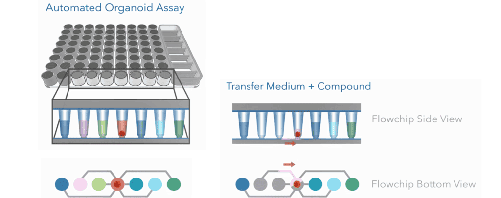
The Pu·MA System 3D with the 3D Flowchip automates assays for 3D cell models using proprietary microfluidic 3D assay technology. This technology allows for multiparametric data collection from a single organoid, spheroid, or tumoroid.
How does it work?
All assay reagents and organoids are loaded into the Pu·MA System 3D Flowchip wells. The flowchips are placed into the the Pu·MA System. Automated reagent exchange takes place through microfluidic channels and controlled by the Pu·MA System.
Types of assay workflows
This technology can enable a range of assay workflows that result in multiparametric readouts from 3D cell models and organoids.
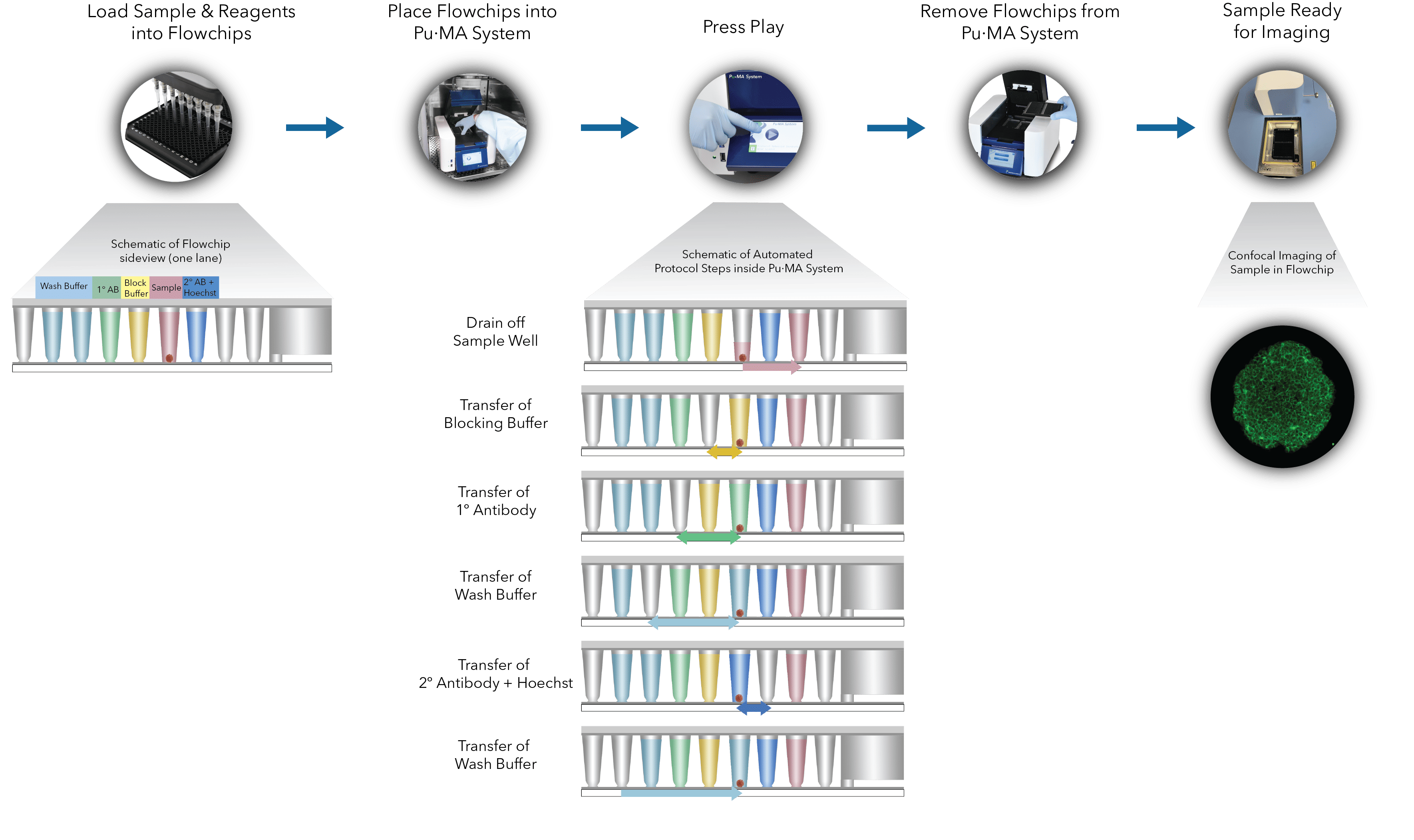
Immunofluorescence for Biomarker Detection
Biomarker detection in organoids, spheroids or tumoroids can be performed using immunofluorescence as shown in the workflow below. Organoids, primary antibody, secondary antibody, washes and nuclear staining are all loaded into the flowchips. Automated steps are performed within the Pu·MA System.
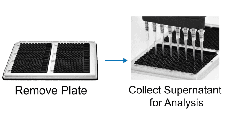
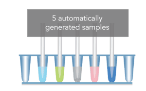
In Situ Supernatant Sampling
Organoids, media and reagents are loaded into the flowchips. The Pu·MA System performs the automated steps described below in the workflow. Read the application note on multiparametric analysis from 3D cell models with compound treatment supernatant sampling supernatant and content imaging.
![]()
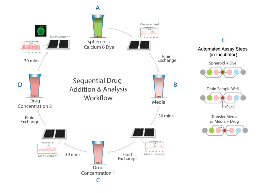
Sequential Drug Addition
Organoids, media and reagents are loaded into the flowchips. The Pu·MA System performs the protocol described below. Different concentrations of drug or compound can be added sequentially via automated steps, during which the samples can be visualized or measurements taken with minimal disruption to the organoid samples. Read the application note on sequential drug addition to neurospheroids to measure calcium oscillations.
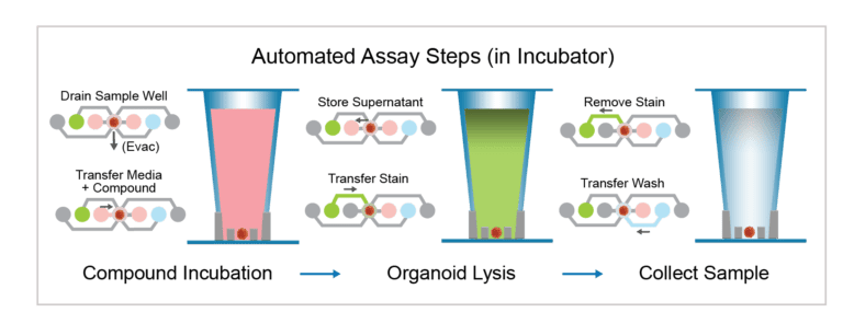
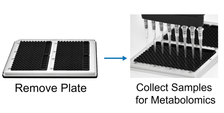
Metabolomics Workflow
Spheroids or organoids, media and metabolites are loaded into the flowchips. The Pu·MA System performs the automated steps described below in the workflow. Once the automated treatment steps are completed, the organoids can be lysed in situ for downstream Mass Spectrometry analysis.
Read about single spheroid metabolomics in the application note.
If you have questions about using the Pu·MA System for applications with your samples, please reach out via the Contact Us page or you can Request a Quote.
Contact Us
Protein Fluidics, Inc.
875 Cowan Road, Suite B,
Burlingame, CA 94010
+1 650 529 5080
info@proteinfluidics.com
#pumasystem #flowchip #3dcellassay
Our Company