Pu·MA System 3D for Automated Sequential Drug Addition Workflow & Analysis for Neurospheroids
Introduction
3D cell models and organoids provide a better representation of in vivo tissue or organ function. For the last three decades researchers have been perfecting the formation and maintenance of various 3D models for understanding both disease and normal physiology (1-3). However, they have been limited by their ability to perform complex assays easily and quickly with these precious samples especially patient-derived material. When manually performing drug treatments and assays in multiwell plates one is often confined to a single readout per sample. For imaging, the manual treatment, staining, and processing of spheroids and organoids is typically labor-intensive and prone to disruption or loss of samples.
The focus of this application note is, the use of our automated Pu·MA System for the sequential drug treatment of neurospheroids for functional evaluation. Neurospheres treated with various neuro-active compounds were analyzed for neuronal activity by Ca2+ oscillations using calcium sensitive dyes and fast kinetic fluorescence imaging. This enabled automated multi-dosing protocols with multiple reagent exchanges for single neurospheroids assay.
Pu·MA System Workflow
Pu·MA System and flowchips have been designed for streamlined workflow as shown in in Figure 1. which consists of:
- loading spheroids and reagents into the flowchips
- placing the flowchips into the Pu·MA System (inside the incubator)
- running automated reagent exchange protocols via an intuitive touchscreen interface
- analyzing the spheroids in the flowchip with systems such as imagers, kinetic fluorescence systems or plate readers
The system architecture and use of pneumatic microfluidics to move fluids provides gas exchange to the sample chambers within the incubator environment.
- a convenient multi-well plate format (384-well spacings SLAS/ANSI standard)
- standard spacing for use with multichannel pipettes or automated liquid dispense system
- up to 32 tests per plate
- optically clear bottoms for imaging with any fluorescence or confocal imaging system
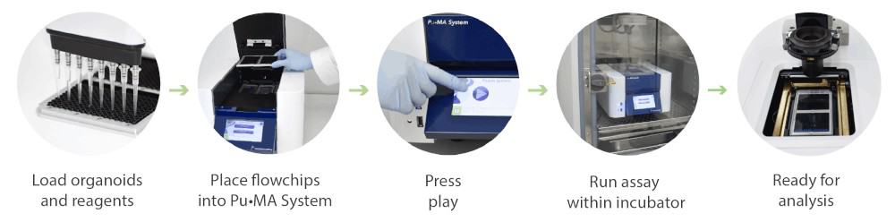
Figure 1. Schematic of the Pu·MA System workflow for automated organoid assays, in situ staining and imaging