Automated 3D co-culture assay with activated T-Cells using the microfluidic Pu·MA System
Introduction
Understanding the tumor – immune cell microenvironment is vital to devising therapeutic interventions. With advances in immunotherapy, 3D models that recapitulate tumor complexity, architecture, cellular interactions, and infiltrating immune populations, become extremely important. 3D models mimic the tumor microenvironment better than a 2D monolayer of cells by simulating the solid tumor mass
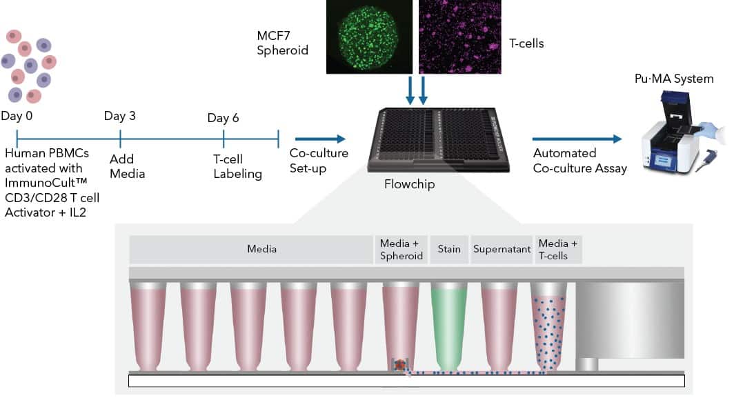
This technical note describes an approach we have developed to perform 3D co-culture assays using the automated microfluidic Pu·MA System®. In this study, we used a co-culture of MCF7 breast cancer spheroids and T-cells from human peripheral blood mononuclear cells (PBMCs). This is an appealing format because it combines a moderately high level of biological complexity, but can have predictable behavior with simpler and reproducible protocols. We analyzed the co-cultures by viability and immunofluorescence (IF) staining. This co-culture platform can be further extended to more complex organoids, patient-derived 3D models, and other immune cell types.

Fig 1. 3D Co-culture workflow
