Assays using Magnetized 3D Cell Models
There are several limiting factors that reduce the ability to perform complex assays easily and quickly, especially with precious patient-derived material. When performing drug treatments and assays in multi-well plates, one is limited by the number of readouts per sample. The manual treatment, staining, and processing of spheroids and organoids is typically labor-intensive and prone to disruption or loss of samples. In addition, high content imaging can be problematic because organoids tend to locate at the edges of wells.
To overcome these issues during assays, we demonstrate the use of our Pu·MA System® 3D MAG and 3D Flowchips to perform automated assay steps with magnetically coated 3D cell models. NanoShuttle™ coated 3D cell models were transferred and centered into flowchip wells using magnets. The cell models were unaffected during the assays and easily imaged using a confocal microscope.
The tabs below showcase this application.
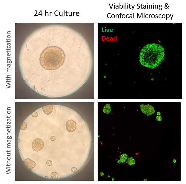
MCF7 Spheroids formed with or without magnetization within Pu·MA System flowchip sample wells. Samples were also analyzed for viability and subsequently imaged using confocal microscope (Yokogawa CellVoyager CQ1)
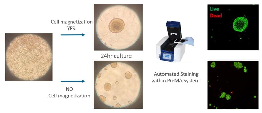
Formation, Magnetization, Transfer & Automated Staining of Organoids
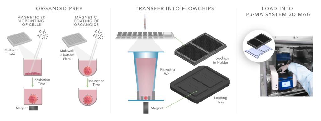
The workflow used 1 μl of NanoShuttle (Greiner Bio-One) per sample to coat the formed 3D cell model. After the incubation they are transferred into flowchips with the aid of the loading tray. Reagents are loaded in the wells adjacent to the sample well and the flowchip holder placed into the Pu·MA System 3D MAG within the incubator.
Improved Assays with Magnetically Coated 3D Cell Models
This application note demonstrates the use of our Pu·MA System® 3D MAG and 3D flowchips to perform automated assay steps with magnetically coated 3D cell models. NanoShuttle™ coated 3D cell models were transferred and centered into flowchip wells using magnets. The cell models were unaffected during the assays and easily imaged using a confocal microscope.
You can learn how you can:
- 20X reduction of pipetting steps = reduced human error
- Zero organoids perturbatoin during multiple assay steps
- Only 1-Touch to run your assay protocol for “hands-off” workflow
- 10X more precise organoid location for microscopy
Automated Assays Using Magnetically Bio-printed 3D Cell Models (2021)
Glauco R. Souza, PhD (Director of Global Business Development & Innovation, Greiner Bio-One)
Evan F. Cromwell, PhD (President & CEO, Protein Fluidics, Inc.)
MCF7 Spheroids formed with or without magnetization for 24 hours within Pu·MA System flowchip sample wells. Samples were also analyzed for viability and subsequently imaged using confocal microscope (Yokogawa CellVoyager CQ1)
Contact Us
Protein Fluidics, Inc.
39655 Eureka Dr
Newark, CA 94560
+1 650 529 5080
info@proteinfluidics.com
#pumasystem #flowchip #3dcellassay
Our Company