Immuno-Oncology & Immune Co-Culture
With advances in disease research, immune-oncology and cancer immunotherapy have rapidly been expanding. Therapeutic approaches such are T-cell therapy and chimeric antigen receptor T-cell therapy (CAR-T), are becoming important approaches for treating cancer with many successful clinical trials. However, researchers are continually discovering new targets and new mechanisms to improve our understanding of the complex tumor microenvironment (TME).
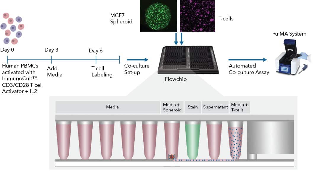
To aid in this field of research, we demonstrate the use of the Pu·MA System for immune-oncology, immune cell co-cultures and cell transfers. Co-cultures can be maintained without disturbing the organoids, you can collect supernatant to analyze secreted factors. You can perform staining steps and image the co-cultures in one simple workflow.
Pu·MA System assays are compatible with:
Sample types: spheroids, patient-derived organoids, tumoroids
Co-culture with: PBMCs, monocytes, macrophages
Sample well can be imaged using confocal or fluorescence microscopy
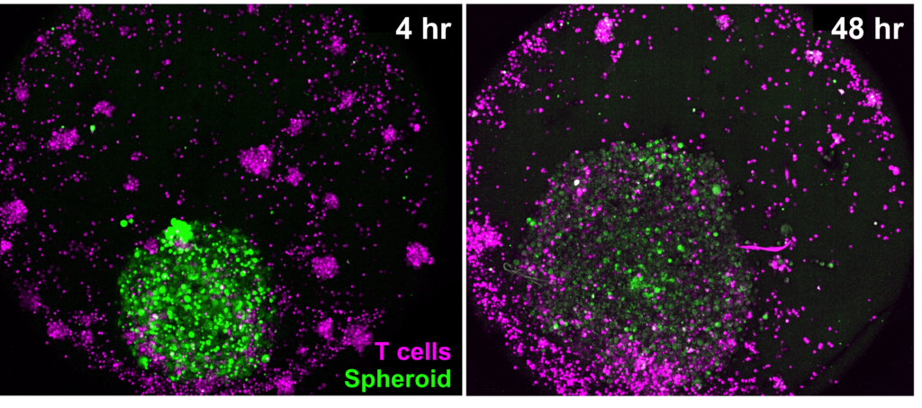
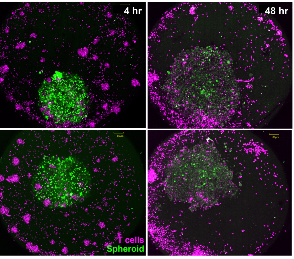
T-cells transferred to MCF7 spheroids in sample wells within Pu·MA System flowchip. The co-culture were analyzed at time 4 hr (at seeding) and after 48 hours of incubation.
In addition, this assay can be customized for collecting supernatants to analyze secreted factors.
Transfer immune cells, perform assays, and image co-cultures
Benefits to your research:
- Zero organoid perturbation during co-culture steps
- Seamless immune cell transfers with no loss of viability
- 5X increased sensitivity for secreted factors in supernatants
Tech Note: Automated co-cultures using spheroids & immune cells
This technical note describes an approach we have developed to perform 3D co-culture assays using the automated microfluidic Pu·MA System®. In this study, we used a co-culture of MCF7 breast cancer spheroids and T-cells from human peripheral blood mononuclear cells (PBMCs). This is an appealing format because it combines a moderately high level of biological complexity, but can have predictable behavior with simpler and reproducible protocols. We analyzed the co-cultures by viability and immunofluorescence (IF) staining.
This co-culture platform can be further extended to more complex organoids, patient-derived 3D models, and other immune cell types.
You can learn how you can:
- 20X reduction of pipetting steps = reduced human error
- Zero organoids perturbation during multiple assay steps
- Only 1-Touch to run your assay protocol for “hands-off” workflow
- 10X more precise organoid location for microscopy
Immuno-Oncology Co-Cultures within the Pu·MA System
Here is some data from T-cells transferred to MCF spheroids for co-cultures and analyzed by confocal microscopy after 48 hours of incubation. MIP of z-stacks acquired on CellVoyager CQ1 Benchtop High-Content Analysis System
Contact Us
Protein Fluidics, Inc.
39655 Eureka Dr
Newark, CA 94560
+1 650 529 5080
info@proteinfluidics.com
#pumasystem #flowchip #3dcellassay
Our Company