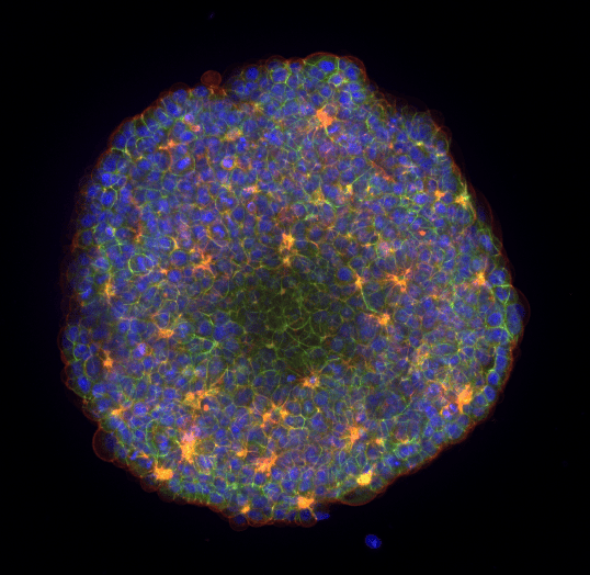
Immunofluorescence Staining for Biomarkers
A key challenge when performing drug or compound testing followed by biomarker detection, is the manual and labor intensive protocol. The immunofluorescence staining workflow has multiple incubation and wash steps which can be additionally complicated when working with precious 3D cell models such as organoid, spheroids or tumoroids.
With the Pu·MA System and flowchips, you can automate your IF staining workflow, make imaging your samples easier, and minimize the costs! Using our innovative protected sample chamber, hands-free fluid transfers, and optically clear COC flowchip bottom, we simplified the IF staining workflow to make getting good data a breeze.
Press Play & Walk Away!

Resources Featuring IF Staining
Automated IF Staining for biomarker detection in organoids and spheroids
- Automate all the IF staining steps
- 20X reduction in pipetting steps compared to manual procedure
- Save over 5 hours of total “hands-on” time
- Samples are ready for high-content imaging within the flowchips
Biomarker Detection in 3D Cell Models
Here is a recording of Dr. Katya Nikolov’s Poster 1173-F which was presented at SLAS2022 in Boston
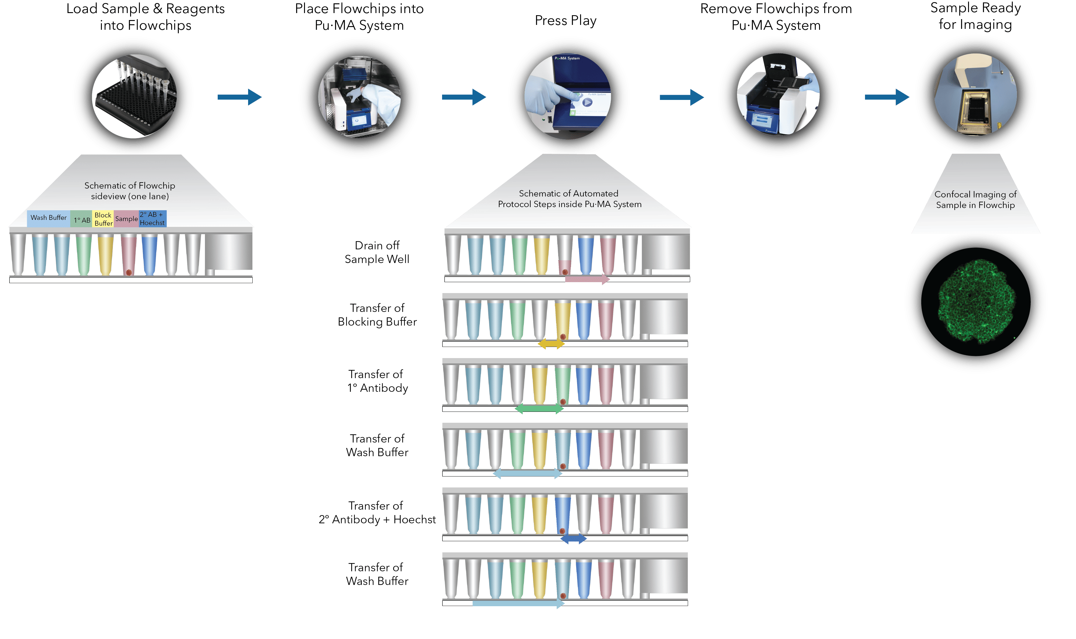
IF Staining Workflow
Organoids, media and reagents are loaded into the flowchips. The Pu·MA System performs the automated steps described below in the workflow or you can download the technote
TECH TALK: Cancer Modeling & High-Content Imaging using 3D Cell-Based Assays
In this two-part webinar we present to you streamlined technologies which can bring consistent timesaving, ease-of-use, and high-quality data to your 3D cell-based workflows:
(A) The Pu·MA System is a microfluidics-based benchtop automated device for performing “hands-off” 3D cell-based assays. In this webinar, we present data from IF staining assays using tumoroids followed by Yokogawa’s high-content imaging systems for biomarker detection.
(B) Yokogawa’s high-content imaging systems such as CellVoyager CQ1 provide superior confocal imaging using the Nipkow Spinning Disk Confocal Technology. In this webinar, details of the high-content imaging capabilities, easy to use and intuitive image acquisition software, especially for increasing productivity and a streamlined workflow are presented.
If you have questions about using the Pu·MA System for applications with your samples, please reach out via the Contact Us page or you can Request a Quote.
Contact Us
Protein Fluidics, Inc.
39655 Eureka Dr
Newark, CA 94560
+1 650 529 5080
info@proteinfluidics.com
#pumasystem #flowchip #3dcellassay
Our Company