Assays using Hydrogels & Other ECM
Organoids and 3D cell models are formed from mammalian tissue or cells. They can be cultured using various methods such as low-attachment multi-well plate formats. But most often specific type of cells do need an extracellular support. Consequently, the formation of many organoids or 3D cell structures requires the support of hydrogels. Accordingly, researchers use scaffolds or hydrogels (either xeno-free chemically defined or complex gels). Depending on the cells or tissue being studied, hydrogels are important for research in drug discovery, compound screening or understanding the biology of the organoids.
In this application page, we demonstrate the use of the Pu·MA System 3D for automating assays with the addition of hydrogels into the flowchip consumables to facilitate work with specialized cells,. You can collect supernatants at multiple time points during the course of an assay without disturbing the spheroid or organoid. You can also perform drug treatment followed by immunofluorescence staining with organoids in hydrogel or Matrigel.
The tabs below showcase this application of assays with the Pu·MA System optimized for use with hydrogels and other ECM.
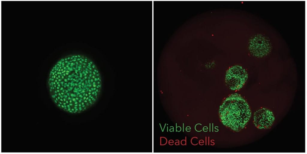
Single or multiple lung organoids in Pu·MA System flowchip sample wells. They were treated with a drug and subsequently stained with Calein AM or ethidium homodimer for viability assessment.
Transfer, Automated Staining, and Imaging of Organoids in Matrigel
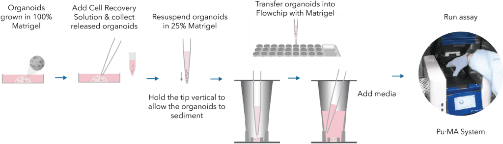
This schematic below shows the workflow for retrieving organoids from a Matrigel dome and transferring them to our flowchips in 25% Matrigel. Subsequently, the flowchips are placed into the Pu·MA System and automated assays can be programed to be executed. A concentration range of 20% to 30% Matrigel was experimentally determined as optimal to support and maintain organoids during automated assays such as IF staining or drug treatments.
App Note: Automated Biomarker Assays for 3D Cell Models on the Pu·MA System® with Xeno-free VitroGel® Hydrogel
In this application note, we demonstrate applications using ECMs and engineered hydrogels for automated image-based profiling 3D assays (Figure 1). We present the use of the Pu·MA System for two 3D cell culture applications using VitroGell
1. Formation of 3D cell models
2. Cell biomarker detection of 3D cell models.
App Note: Automated Assays with 3D Cell Models with ECM
To streamline complex multi-step assays and data acquisition from 3D cell models in ECM, we have developed and automated workflows for Pu·MA System. In this app note, we present three different case studies which demonstrates the efficiency and performance of the Pu·MA System for assays with organoids and spheroids.
- Formation of 3D cell models in ECM
- Cell marker detection of 3D cell models in ECM
- Profiling engineered alveolar organoids in hydrogel
Tech Note: Transfer, Automated Staining, and Imaging of Organoids in Matrigel
This technical note describes the procedure of retrieving pulmonary organoids from a Matrigel dome and transferring them to a Flowchip in 25% Matrigel to perform automated assays using Pu·MA System. A concentration range of 20% to 30% Matrigel was experimentally determined as optimal to support and maintain organoids inside the protected chamber of the flowchip.
You can learn how you can:
- 20X reduction of pipetting steps = reduced human error
- Zero organoids perturbation during multiple assay steps
- Only 1-Touch to run your assay protocol for “hands-off” workflow
- 10X more precise organoid location for microscopy
Biomarker Detection in 3D Cell Models
Here is a recording of Dr. Katya Nikolov’s Poster 1173-F which was presented at SLAS2022 in Boston. This poster includes workflows involving hydrogels such as Matrigel or VitroGel.
Patient-Derived Tumoroids in Hydrogel with Automated Biomarker Detection
In this two-part webinar we share with you:
(A) VitroGel®, a xeno-free, bio-functional hydrogel system for 3D cell culture applications. The state-of-the-art hydrogel system from TheWell Bioscience gives an outstanding balance of biological functions and operating ease to establish 3D cell models. We will introduce the robust features of our 3D platform with multiple culture method including 3D cell culture, 2D hydrogel coating, static suspension culture, hydrogel-cell bead, and as an injectable carrier. The ready-to-use VitroGel Hydrogel Matrix will be presented as an example for tumoroid long-term culture and co-culture models.
(B) Pu·MA System, a benchtop automation device for performing 3D cell-based assays. In this webinar, we present data using triple negative breast cancer samples in xeno-free VitroGel for biomarker detection upon drug treatment within the microfluidics-based Pu·MA System. Subsequently, the integrated workflow is followed by confocal microscopy of the assay flowchips. Further we show that for these patient-derived samples, we observed no phenotypic differences when compared to Matrigel but found better ease of use with VitroGel such as temperature-independent and tunable gel properties.
Contact Us
Protein Fluidics, Inc.
39655 Eureka Dr
Newark, CA 94560
+1 650 529 5080
info@proteinfluidics.com
#pumasystem #flowchip #3dcellassay
Our Company