Webinar Recording
Automated 3D Cell-Based Assays Using a Novel Flowchip System
Evan F. Cromwell, PhD (President & CEO, Protein Fluidics, Inc.)
Mar 31, 2021
Summary
There is an increasing interest in using three-dimensional (3D) cell structures for modeling tumors, organs, and tissue to accelerate translation research. Significant progress has been made in formation of such structures to recapitulate the in vivo environment but performing complex assays with them can be challenging. Organoids can be subject to loss or damage from pipettes during common steps such as media exchange, supernatant sampling, or immunofluorescence staining.
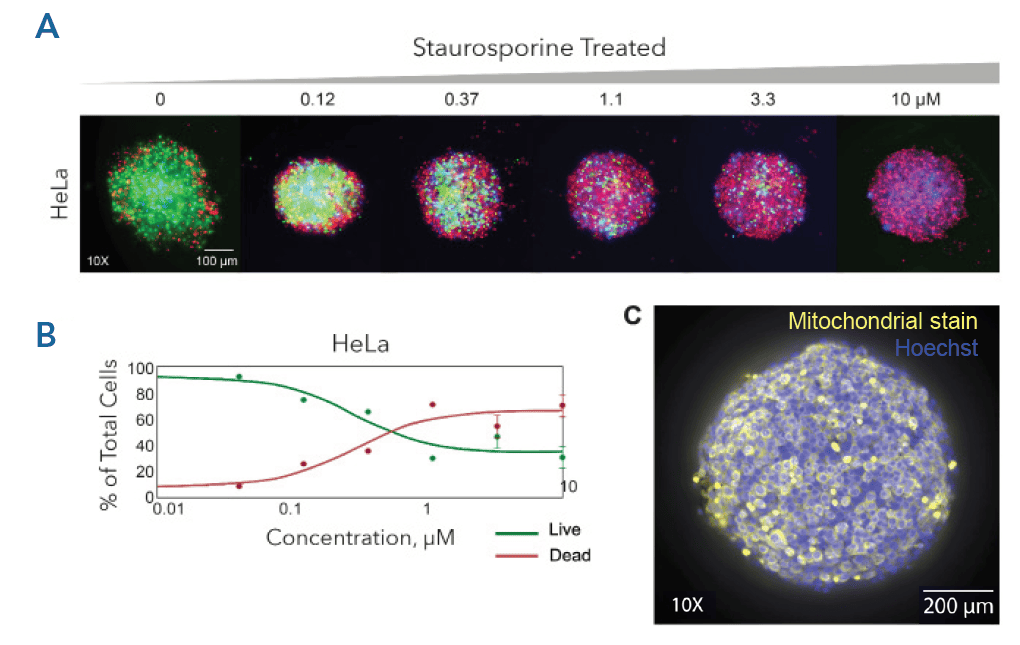
We presented results from automated organoid assays using a Pu·MA System for single organoid secretion, and metabolite sampling. This webinar featured different assays using different types of 3D cell models.
Learn How to
- Automate media exchange and staining of 3D models such as spheroids, organoids, and tumoroids
- Acquire high-content images within the flowchips without disturbing the
3D cell models - Collect multiple supernatants during the assay to study phenotypic changes and compound effects
- Perform multifunctional profiling from single samples

Contact Us
Protein Fluidics, Inc.
875 Cowan Road, Suite B,
Burlingame, CA 94010
+1 650 529 5080
info@proteinfluidics.com
#pumasystem #flowchip #3dcellassay
Our Company
