Tumoroid Toxicity Testing and Imaging
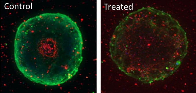
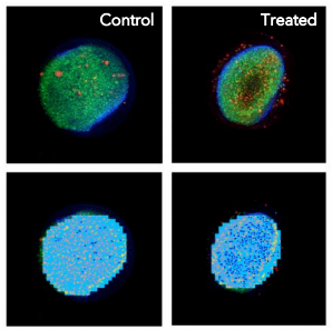
Spheroids and other 3D cell models are routinely used for toxicity testing and screening. With the Pu·MA System 3D, you can perform automated drug treatments without disturbing the organoid.
Automated drug treatments are performed during the course of the assay, followed by transfer of staining solution, and then confocal imaging within the flowchips.
The tabs below showcase this application of toxicity testing and imaging using the Pu·MA System.

Pu·MA System 3D for Spheroid Toxicity Testing and In Situ Imaging

This application note demonstrates the use of the Pu·MA System for spheroid treatment with drugs followed by in situ lysis for preparing samples for downstream metabolomics analysis.

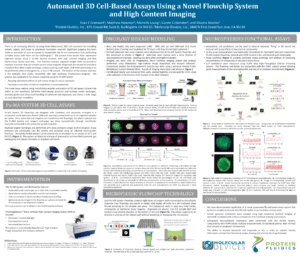
Automated Long-Term 3D Cell-Based Toxicity Studies Using a Flowchip System
We presented data about how you can single spheroid drug treatment and dosing. The spheroid samples were lysed within the incubator and samples analyzed for metabolic changes.
Toxicity Testing and Imaging Workflow
Organoids, media and reagents are loaded into the flowchips. The Pu·MA System performs the automated steps described below in the workflow.

Automated 3D Cell-Based Assays Using the Pu·MA System
In this webinar we discuss automated 3D cell-based assay and how you can perform single spheroid drug treatment and dosing.
Learn How to
- Automate media exchange and staining of 3D models such as spheroids, organoids, and tumoroids
- Acquire high-content images within the flowchips without disturbing the
3D cell models - Collect multiple supernatants during the assay to study phenotypic changes and compound effects
- Perform multifunctional profiling from single samples
Contact Us
Protein Fluidics, Inc.
26219 Eden Landing Rd
Hayward, CA, 94545
+1 650 529 5080
info@proteinfluidics.com
#pumasystem #flowchip #3dcellassay
Our Company
